Sunday, November 18, 2007

Final Week of Observation

This week I snagged some photos for all of you avid readers of this blog (not that there are any). However, I did get photos of the life forms I have previously talked about. This week in pictures:

NOSTOC


Here we have some pretty good pictures of Nostoc. Higher order taxonomy: Eubacteria, Cyanobacteria, Nostocales, Nostcaceae. Nostoc is the genus, and is composed of several species of gelatinous colonies of filaments (the trichomes we've talked about previously) surrounded by a thin "sheath." Nostoc are nitrogen-fixing and photosynthetic (but do not have chloroplasts).
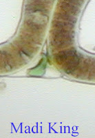
SCYTONEMA


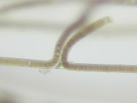
Above you can see some spectacular pictures of Scytonema, a cyanobacteria. Many details of this cyanobacteria are included in previous posts, so I won't drag on about it. However, some cool pictures of Sytonema in real life places can be seen at the first website. Plus, a fantastic picture can be found at the second (note the clarity!).
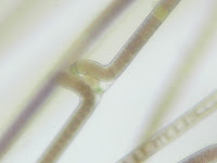
ANKISTRODESMUS


Above you can see the Ankistrodemus. Each line of the "jack" like formation is actually a seperate specimen. As you can tell, each one is long and needle-shaped. The Ankistrodesmus is very common in North America in freshwater ponds and lakes, and can even be found in waterfalls. This species has a high tolerance for copper treatments, which are commonly used to control algal growth.

OTHER ALGAE


Various other forms of green life are depicted above.
CHAETOCHASTER DIASTOPHUS
The above life form is related to the earthworm. This is the worm I refer to in earlier posts.

Sunday, November 11, 2007

Week Four

This week in the biology lab, I found another cyanobacteria to be present amidst all the Nostoc. I was lucky enough to see some "Scyntonema." This cyanobacteria comes from the Scyntonemataceae family. I have found two online pictures to depict this cyanobacteria's famous feature of false-branching. In these pictures you can see how two trichomes meet and seeming grow into each other. Also, in the second picture you can see the heterocysts present in the cyanobacteria. The first two links go with the pictures, respectively. The final link has some interesting information about nitrogen-fixation and about lichens.


http://www.msu.edu/course/bot/423/AlgalMaster.html
http://vis-pc.plantbio.ohiou.edu/algaeimage/pages/Scytonema.html
http://www.biology.duke.edu/bio265/Archive/1999/czcf/scytonema.html

*I added a little bit of water to the microcosm this week.*

Sunday, November 4, 2007

Week Three

This week showed an explosion of green in my microcosm. One kind of cyanobacteria in particular seems to have taken over the habitat. Nostoc, one of the cyanobacteria we looked at in lab unit 13, has grown to a raging population over the past few weeks. Nostoc is a freshwater cyanobacteria (obviously) that have long chains of cells known as filaments. Nostoc contains photosynthetic pigments.
Also, since I last visited my microcosm, one pellet of "Atison's Betta Food" was added. This may have played a factor in the sudden cyanobacteria growth I observed.

http://en.wikipedia.org/wiki/Nostoc

Saturday, October 27, 2007

Week Two

I visited my micrcosm on Wednesday. This week, to my surprise, the aquarium was overflowing with life. The life form I spent the most time observing, was a worm like creature. I spotted two of these swimming around in the aquarium and followed them both for quite some time. The organism was clear, long, and covered in cilia. I found these guys to love a diet of moss. While I was watching one of them, he consumed an amazing amount of moss for such a little guy. I was able to actually see the moss moving through his "digestive track." Unfortunately, I was only able to stay for an hour and a half, and did not have time to get any photos of these creatures. Hopefully next time I'll be able to share some images of them with you!

Sunday, October 21, 2007

Initial Observations

The first time I checked the MicroAquarium for signs of life, there was scarce to see. There were three organisms that I did notice.
The first organism was stationary, and chlorophyll green. It was located near the bottom of the aquarium, just above some mud. Based upon these things, the organism was recognized as green algea. This was the only green algea I found.
The second organism that I saw was motile. It appeared to be rounded and its head was a seperate segment. It looked very similar to a very small ladybug that you could see through. It also appeared to have two very faint tails that forked out from its body. The organisms appeared to wiggle back and forth, using its two tails like a fin. This was the only one of its kind that I saw.
Finally, I noted a worm-like organism that also had a tail. This organism's tail was very short. The organism spent most of its time spinning in circles, and occasionally moving forward. This organism was rather hard to keep up with under the microscope, and was the only one of its kind that I was fortunate enough to see.
And so concludes the rather short list of living things I observed on the first day. Better luck next time, I hope.

The Beginning

The building of my microcosm began with a MicroAquarium kit. The kit includes a glass tank, a stand and a lid. From the water sample provided, I filled 1/3 of the MicroAquarium with water from the bottom of sample one. This water also had mud in it. I filled another 1/3 of the tank with water from the middle of sample one. Finally, I filled the remainder of the tank with water obtained by skimming the top of sample one. The water in sample one was originally obtained from a loow water pool in a side channel of the Pigeon River. This particular channel can be found near Harmon Road, which can be found off of the Harmon Dean Exit of I 40. This particular part of the interstate is located in Haywood County, North Carolina. The water in the part of the channel sampled is reported to recieve full sun exposure.
After filling the MicroAquarium with the water sample, I added about a one inch piece of moss. This particular moss is known as Rhynchostegium serrulatum, and was collected from the greenhouse in the White Ave Biology Annex, which is located at The University of Tennessee. This moss was grown in partial shade exposure.